
On this page
The basic ABG interpretation steps are: check the pH, find the primary problem by comparing PaCO2 and HCO3−, decide whether the body is compensating, and then look at oxygenation. Work through the same order every time and most arterial blood gas questions, on the ward or in the NCLEX and Philippine board exams, become quick to solve.
Key points
- Typical adult ranges: pH 7.35–7.45, PaCO2 35–45 mm Hg, HCO3− 22–26 mEq/L, PaO2 75–100 mm Hg. Ranges vary by laboratory.
- PaCO2 is the respiratory (lung) value; HCO3− is the metabolic (kidney) value.
- The value that moves in the direction of the pH problem is the primary disorder; the other one is compensating.
- A normal pH does not always mean a normal ABG: look for full compensation or a mixed disorder.
What is an arterial blood gas (ABG)?
An ABG is a blood test taken from an artery, usually the radial artery at the wrist, though the brachial and femoral arteries can also be used. It shows how well the lungs move oxygen in and carbon dioxide out, and whether the blood is too acidic or too alkaline.
Abnormal results can come from lung disease, kidney disease, metabolic problems such as diabetic ketoacidosis, medicines, or injuries that affect breathing. Venous blood gases are sometimes used instead, but venous values differ: venous PO2 is much lower and venous pH slightly lower than arterial.
ABG normal values
| Value | Typical adult range | What it tells you |
|---|---|---|
| pH | 7.35–7.45 | Acid–base status: below 7.35 = acidemia, above 7.45 = alkalemia |
| PaCO2 | 35–45 mm Hg | Respiratory component (ventilation) |
| HCO3− | 22–26 mEq/L | Metabolic component (kidneys, buffers) |
| PaO2 | 75–100 mm Hg | Oxygenation |
| SaO2 | 95–100% | Oxygen saturation of haemoglobin |
| Base excess | −4 to +2 | Metabolic component (extra check) |
Oxygen values are lower in people living at higher altitude, so interpret PaO2 with the patient’s setting and oxygen therapy in mind.
ABG interpretation steps
Step 1: Look at the pH
Below 7.35 is acidemia; above 7.45 is alkalemia. If the pH is within the normal range, use 7.40 as the midpoint: 7.36–7.39 leans acidic and 7.41–7.44 leans alkaline. This tells you which way the primary problem points.
Step 2: Find the primary disorder
Check which value matches the pH. If PaCO2 is high with a low pH, or low with a high pH, the cause is respiratory. If HCO3− moves in the same direction as the pH (low with acidosis, high with alkalosis), the cause is metabolic.
Step 3: Check for compensation
Now look at the value that did not cause the problem. If it is normal, the disorder is uncompensated. If it has moved in the opposite direction to the primary problem and the pH is still abnormal, it is partially compensated. If both values are abnormal but the pH is back in the normal range, it is fully compensated.
Step 4: Assess oxygenation
Look at PaO2 and SaO2 for hypoxemia, taking into account how much oxygen the patient is receiving.
Step 5: For metabolic acidosis, go further
Calculate the anion gap: Na+ − (Cl− + HCO3−). A recent review gives 8–16 as normal, with higher values pointing to added acids such as ketones or lactate; reference values differ between laboratories. To check whether breathing is compensating as expected, use Winter’s formula: expected PaCO2 = (1.5 × HCO3−) + 8 ± 2. A measured PaCO2 outside that range suggests a second, mixed disorder.
The four acid–base disorders at a glance
| Disorder | pH | Primary change | Compensation | Common causes |
|---|---|---|---|---|
| Respiratory acidosis | ↓ | PaCO2 ↑ | HCO3− ↑ | Hypoventilation (slow or shallow breathing), respiratory failure |
| Respiratory alkalosis | ↑ | PaCO2 ↓ | HCO3− ↓ | Hyperventilation, for example from anxiety |
| Metabolic acidosis | ↓ | HCO3− ↓ | PaCO2 ↓ | DKA, septic shock, kidney failure, toxins, bicarbonate loss from the gut |
| Metabolic alkalosis | ↑ | HCO3− ↑ | PaCO2 ↑ | Prolonged vomiting, diuretics, hypokalemia, hypovolemia |
Metabolic (kidney) compensation takes days to become effective, so a respiratory problem with strong metabolic compensation is usually not brand new. Electrolytes matter here too: low potassium and metabolic alkalosis often appear together, so review our electrolytes guide for nurses alongside this one.
Practice examples
- pH 7.30, PaCO2 55, HCO3− 25. Acidemia; PaCO2 is high and matches; HCO3− is normal. Answer: uncompensated respiratory acidosis.
- pH 7.32, PaCO2 30, HCO3− 15. Acidemia; HCO3− is low and matches; PaCO2 is low (compensating) but pH is still abnormal. Answer: partially compensated metabolic acidosis. Winter’s formula: 1.5 × 15 + 8 = 30.5 (range 28.5–32.5), so compensation is appropriate.
- pH 7.43, PaCO2 50, HCO3− 32. pH normal but above 7.40; HCO3− is high and matches; PaCO2 is high (compensating). Answer: fully compensated metabolic alkalosis.
Nursing assessment
- Respiratory rate, depth and effort; level of consciousness; skin colour; SpO2.
- Current oxygen device and flow or FiO2. MedlinePlus advises that oxygen settings stay constant for 20 minutes before an ABG is drawn.
- Anticoagulant or antiplatelet use, including aspirin, which raises bleeding risk at the puncture site.
- Circulation to the hand before a radial puncture (see below).
- Related labs: electrolytes, glucose, lactate and ketones when metabolic acidosis is suspected.
Nursing considerations when collecting an ABG
- Modified Allen test: occlude both the radial and ulnar arteries while the patient clenches the fist, then release the fist and the ulnar pressure. Colour returning within about 10–15 seconds suggests adequate collateral flow; if it does not, the radial site should not be used.
- Use a heparinised blood gas syringe, fill it fully, and remove air bubbles at once. Air in the sample can falsely lower PCO2 and raise pH and PO2.
- Mix gently, label with the oxygen therapy details, and send for analysis promptly, on ice if your facility requires it.
- Apply firm pressure to the site after the needle is removed and watch for hematoma, bleeding and changes in circulation to the hand. Follow your facility’s policy on duration.
- Report critical values and changes in the patient’s condition promptly.
Exam tips
- ROME: Respiratory = Opposite (pH and PaCO2 move in opposite directions); Metabolic = Equal (pH and HCO3− move the same way).
- Trap: a normal pH with abnormal PaCO2 and HCO3− is full compensation, not a normal result. Use 7.40 to decide which side it leans.
- Classic pairs: prolonged vomiting or diuretics → metabolic alkalosis; DKA or bicarbonate loss from the gut → metabolic acidosis; hypoventilation → respiratory acidosis; anxiety with hyperventilation → respiratory alkalosis.
- Many students find the “tic-tac-toe” method helpful: label each value acid, normal or base, and see which two line up.
Frequently asked questions
What is the fastest way to interpret an ABG?
Use the same three questions every time. Is the pH acidic or alkaline? Which value, PaCO2 or HCO3−, moves with that pH? Is the other value normal, moving to compensate, or bringing the pH back to normal? Then check PaO2. With practice this takes under a minute.
Why is HCO3 metabolic and PaCO2 respiratory?
Carbon dioxide is removed by breathing, so PaCO2 rises with hypoventilation and falls with hyperventilation. Bicarbonate is a base regulated mainly by the kidneys and lost or gained through metabolic processes such as diarrhoea, vomiting or kidney disease. That is why each value points to a different body system.
Is a normal pH always a normal ABG?
No. When compensation is complete, the pH can sit inside 7.35–7.45 even though PaCO2 and HCO3− are both clearly abnormal. A mixed disorder can also cancel out at the pH level. Always look at all the values together with the patient’s condition and history.
Related on Nurses Notes
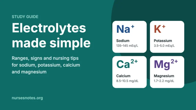
- Electrolytes made simple: sodium, potassium, calcium and magnesium for nurses
- Signs of dehydration in adults and what to do
- All nursing study guides
Sources
- Castro D, Zubair M. Arterial Blood Gas Analysis: Fundamentals, Interpretation, and Clinical Utility. StatPearls, NCBI Bookshelf, updated 2026.
- MedlinePlus Medical Encyclopedia. Blood gases. U.S. National Library of Medicine, reviewed 2024.
- Núñez Sanagustín M, Osredkar J. Blood gas analysis: clinical applications, interpretation and future directions. Medicine International, 2025.
This article is for education only and does not replace advice from your own doctor, nurse or other qualified health professional. Always follow your facility’s policies and reference ranges.